
An OCT eye scan is a non-invasive 3D scan of the structures at the back of the eye. OCT stands for optical coherence tomography and allows an optometrist to see detailed cross-sectional images of the retina and optic nerve.
Sometimes described as a 3D eye scan, OCT provides information that cannot be obtained from a standard retinal photograph alone. It is an important diagnostic tool used by optometrists to assess eye health and detect, monitor and manage a range of eye conditions.
At Rose Optometry in Berry, we use OCT imaging as part of our comprehensive approach to eye health, helping us assess the retina, macula and optic nerve in greater detail.
What is an OCT eye scan?
OCT uses light to create highly detailed images of the tissues inside the eye. The scan produces cross-sectional images that allow your optometrist to examine the different layers of the retina and the optic nerve.
Unlike a conventional photograph of the back of the eye, which provides a two-dimensional image, an OCT scan allows us to look through the layers of the retina.
This makes OCT particularly useful for identifying subtle changes that may not be obvious during a routine eye examination.
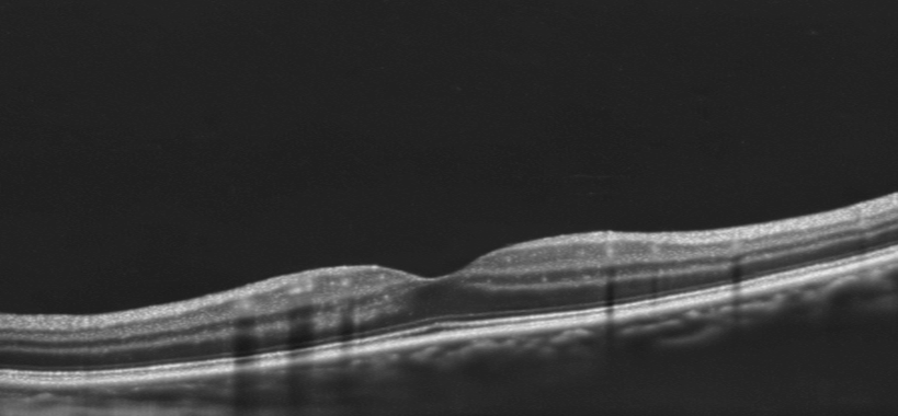
Cross section image of the macula using OCT. Different layers of the retina can be observed by your optometrist.
What does an OCT scan show?
An OCT scan can provide detailed information about structures including:
The retina and its individual layers
The macula, which is responsible for detailed central vision
The optic nerve
The retinal nerve fibre layer
Changes in the thickness or structure of retinal tissue
Fluid or swelling within or underneath the retina
OCT scans can also be compared with previous scans over time. This is particularly valuable when monitoring conditions where gradual changes may occur.
Why do optometrists use OCT?
OCT is used to help detect, assess and monitor eye disease.
It can be particularly useful when assessing conditions such as:
Glaucoma
Glaucoma can damage the optic nerve and cause progressive loss of vision. OCT can measure structures around the optic nerve, including the retinal nerve fibre layer, helping optometrists identify changes that may be associated with glaucoma.
OCT is one part of a glaucoma assessment and is considered alongside other information such as eye pressure, visual field testing and examination of the optic nerve.
Age-related macular degeneration
The macula is the central part of the retina responsible for detailed vision. OCT provides detailed images of the macula and can help identify structural changes associated with age-related macular degeneration (AMD).
It can also help identify changes such as fluid or swelling that may require further investigation or management.
Diabetic eye disease
Diabetes can affect the blood vessels and tissues of the retina. OCT can provide detailed information about the retinal structure and can help identify changes associated with diabetic eye disease, including retinal swelling.
Macular conditions
OCT is particularly valuable when assessing changes affecting the macula. It can help identify abnormalities that may not be obvious from a standard retinal photograph.
Is an OCT eye test the same as a regular eye test?
No. An OCT eye test is not a replacement for a comprehensive eye examination.
A comprehensive eye examination assesses many aspects of your vision and eye health, including visual acuity, prescription, eye movements, eye pressure, the front of the eye, retina and optic nerve.
OCT adds another level of information by providing detailed images of the retinal and optic nerve structures.
Think of it as another tool that allows your optometrist to examine your eyes in greater detail.
What happens during an OCT scan?
An OCT scan is quick, painless and non-invasive.
You simply look at a target inside the OCT instrument while the machine captures detailed images of your eye. There is no contact with the eye and the scan does not involve radiation.
Your optometrist can then analyse the images and look for any changes or abnormalities.
In many cases, an OCT scan takes only a few minutes.
Who should have an OCT eye scan?
OCT can be particularly useful for people who:
Have glaucoma or are at increased risk of glaucoma
Have a family history of certain eye conditions
Have age-related changes affecting the macula
Have diabetes
Have unexplained changes in vision
Are being monitored for an existing eye condition
Have findings during an eye examination that require further investigation
Need detailed monitoring of the retina or optic nerve
Whether an OCT scan is appropriate depends on your individual circumstances. Your optometrist can determine whether OCT imaging would be beneficial as part of your eye examination.
Can an OCT scan detect eye disease before I notice symptoms?
In some cases, yes.
One of the advantages of OCT is that it can reveal structural changes in the retina or optic nerve that may occur before noticeable changes in vision.
This is one reason regular eye examinations are important. Some eye conditions can develop gradually without obvious symptoms in their early stages.
OCT can also provide a baseline image that can be compared with future scans, making it easier to identify changes over time.
Why are OCT scans useful for monitoring eye health?
An individual OCT scan provides valuable information, but repeated scans can be particularly useful.
When your optometrist has previous OCT images available, they can compare your current scan with earlier examinations. This can help determine whether a finding is stable or whether there has been a change.
For conditions such as glaucoma and macular disease, monitoring changes over time can be an important part of ongoing eye care.
OCT eye scans at Rose Optometry
At Rose Optometry in Berry, we use advanced diagnostic technology to provide detailed assessments of both vision and eye health.
Our practice has OCT imaging and wide-field retinal imaging, allowing us to examine different aspects of the eye in detail.
OCT provides a detailed 3D view of the retinal and optic nerve structures, while wide-field retinal imaging provides a broader view of the back of the eye.
These technologies are used alongside a comprehensive clinical examination and other appropriate testing. They help us detect changes, investigate symptoms and monitor eye health over time.
Book an eye test in Berry
If you are due for an eye examination, or if you have concerns about changes in your vision or eye health, an optometrist can determine whether an OCT eye scan is appropriate for you.
At Rose Optometry, we provide comprehensive eye examinations and advanced retinal imaging for patients in Berry and the surrounding Shoalhaven region, including Nowra, Bomaderry, Gerringong, Kiama and Kangaroo Valley.
An OCT scan is more than just a picture of your eye — it gives your optometrist detailed information about the structures that are important for healthy vision.
